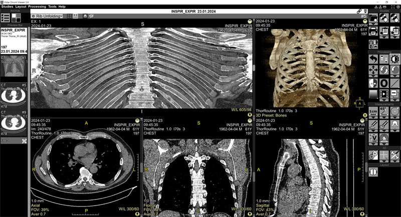
Vidar Dicom Viewer 3.4.2

New version of Vidar Dicom Viewer has been released
The update includes:
New Program
Ribs Unfolding
Program Improvements
- a context menu has been added on the right-click on the image for quick access to the most frequently used functions of the program
- Ultrasound: support for measurements based on region
- visceral fat - added abbreviation explanation from the table
- local archive auto-cleaning
- selection of image type while saving to a file (jpg, bmp, png, tiff)
- improved study filter for Q/R window
Program Fixes
- fixed overlay cropping for Windows 7 systems
- fixed text cropping in volume drop-down list in series header for multi-volume
- fixed definition of some CT-ortho as volumes
- Q/R, fixed error in filtering text with Cyrillic
- fixed crash of CT Perfusion Program due to incorrect definition of block time
- fixed error in spine marking for volume with odd width
- fixed image decompression error
- fixed shutdown when OS is turned off
- 3D export to STL - fixed errors:
- threshold setting via WL with mouse on 3D did not change values in the dialog box
- 3D smoothing filter was saved after export
- “Knife” tool returned
- initially applied median filter on 3D
- fixed authentication problems when importing studies from PAKS via DICOMweb protocol
- fixed bugs of skull removal in CT Perfusion
- improved reading of CT Perfusion studies from Toshiba
- fixed merging of Tomosynthesis series from Siemens
- support for MRI data from Anke devices: merging of diffusion series, added the ability to visualize physically impossible unreliable negative ICD values, as on the diffusion map transmitted from the device
 VDViewer